Pages

Showing posts with label cardiology. Show all posts

Management of atrial fibrillation (AF)

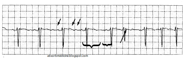
Atrial fibrillation. The tracing demonstrates the absence of P waves (long arrow), as well as the presence of the fine f waves of atrial fibrillation (short arrows). Note the irregularity of the ventricular response, as seen from the variable R-R interval (brackets). [Source]
Common cardiac and non-cardiac causes of aftial fibrillation.

It is not always possible to restore and maintain sinus rhythm in patients with AF.

If sinus rhythm cannot be maintained, treatment should be directed towards controlling the heart rate with
  • digoxin, 
  • ß-blockers, 
  • rate-limiting calcium-channel blockers (verapamil or diltiazem) or 
  • amiodarone. 

Algorithm for the treatment of diastolic and systolic heart failure

Algorithm for the treatment of diastolic or systolic dysfunction. (ACE = angiotensin-converting enzyme; NYHA = New York Heart Association; IV = intravenous.) [Source]

Pulsus Alternans

Pulsus Alternans. [Source]

Pulsus alternans is found in patients with acute left ventricular failure. Alternate weak and strong pulses occur, which are regularly placed. A pathological third heart sound is usually associated. The condition may be associated with heart failure or pericarditis for example, but is not usually seen in association with pericardial effusion.

Signs of complete heart block

The atrial rate is approximately 100 bpm. The ventricular rate is approximately 40 bpm. The two rates are independent; there is no evidence that any of the atrial impulses are conducted to the ventricles. [Source]
Two ECG strips obtained in the same patient simultaneously (each represents a different vector of the heart’s electrical conduction), demonstrating complete AV block (also called 3rd degree heart block). The solid arrows point to P waves, representing atrial electrical conduction originating from the sinus node. The dashed arrows denote electrical conduction in the ventricles (QRS complexes). Note that the P waves are not related to the QRS complexes, demonstrating that the atria are electrically disconnected from the ventricles. The QRS complexes represent an escape rhythm arising from the ventricle. [Source]

Causes of pulmonary hypertension

Pulmonary Hypertension [Source]
Pulmonary hypertension is the narrowing of the pulmonary arterioles within the lung. The narrowing of the arteries creates resistance and an increased work load for the heart. The heart becomes enlarged from pumping blood against the resistance.

Some symptoms include chest pain, weakness, shortness of breath, and fatigue.

The goal of treatment is control of the symptoms, although the disease usually develops into congestive heart failure.

Causes of dilated cardiomyopathy (DCM)

Dilated Cardiomyopathy. [Source]
Dilated cardiomyopathy encompasses a heterogeneous group of conditions.
This is the most common type of cardiomyopathy.

In this disorder, your heart's main pumping chamber — the left ventricle — becomes enlarged (dilated), its pumping ability becomes less forceful, and blood doesn't flow as easily through the heart.

Although this type can affect people of all ages, it occurs most often in middle-aged people and is more likely to affect men. Some people with dilated cardiomyopathy have a family history of the condition. About 25% of cases are inherited as an autosomal-dominant trait.

Treatment of malignant hypertension


An emergency hypertensive is evidenced by markedly elevated blood pressure and target organ damage. This situation requires immediate attention to prevent disability or death. Here, the treatment aim is to reduce the blood pressure promptly but partially to prevent end-organ damage without compromising tissue perfusion. The initial target is to lower the mean arterial pressure (MAP) by no more than 25%, or reduce the diastolic blood pressure by one-third.

AHA 2010 ACLS Algorithm


American Heart Association 2010 ACLS Algorithm.

Defibrillation for adult ventricular fibrillation


Ventricular Fibrillation. [Source]
Three-quarters of arrests are due to ventricular fibrillation. Only a small proportion is due to electromechanical dissociation (EMD), the rest being due to asystole.

EMD usually has a potentially reversible cause:
  • hypovolaemia
  • hypoxia
  • hyperkalaemia
  • hypokalaemia
  • hypothermia
  • tension pneumothorax
  • tamponade
  • toxicity due to drugs
  • thromboembolism

Initial treatment of unstable angina (UA)

The initial treatment of  unstable angina (UA) should include
  • bed rest, 
  • anti-platelet therapy,
  • anticoagulation and 
  • a ß-blocker. 
Algorithm for risk stratification and treatment of patients with UA/NSTEMI. DM= diabetes mellitus; Rx = treatment. Updated with permission from Braunwald E, Zipes DP, Libby P, eds. Heart Disease: A Textbook of Cardiovascular Medicine. 6th ed. Philadelphia, Pa: W.B. Saunders; 2001:1232–1263.[Source]


HCM: A disease of energy deficiency

HCM: A disease of energy deficiency. ADP = adenosine diphosphate; AMP = adenosine monophosphate; ATP = adenosine triphospate; Cr = creatine; FAM = fatty acid metabolism. [Source]
As indicated in red, the phenotype of hypertrophic cardiomyopathy (HCM) can arise from:

Sarcomere disruption in hypertrophic cardiomyopathy (HCM)

Sarcomere disruption in hypertrophic cardiomyopathy (HCM).
ACE = angiotensin-converting enzyme.
[Source]
Hypertrophic cardiomyopathy (HCM) is thought to occur primarily through sarcomere disruption in the face of intact cytoskeleton/sarcolemma.

Risk factors for sudden death in hypertrophic cardiomyopathy (HCM)

Hypetrophic Cardiomyopathy [Source]
Hypertrophic cardiomyopathy (HCM) is the commonest form of cardiomyopathy, with a prevalence 100 per 100,000.

Genetic
It is a genetic disorder with autosomal-dominant transmission, a high degree of penetrance and variable expression.

Clinical features
Symptoms and signs are similar to those of aortic stenosis, except that the character of the pulse is jerky in HCM.


Common organisms in infective endocarditis (IE)


Infective endocarditis on native valves – prevalence of organisms:
This cross-sectional view shows vegetations (accumulations of bacteria and blood clots) on the four valves of the heart. [Source]

  • Streptococci 
    • Viridans group 10–15%
    • Enterococci 20–25%
    • Other
  • Staphylococci 
    • Staphylococcus aureus 9–27%
    • Coagulase-negative 1–3%
  • Gram-negative bacilli
    • Haemophilus spp. 3–8%
  • Anaerobes less than
    • Rickettsia/fungi 2%

Postpartum hypercoagulability and prophylaxis anticoagulants

Blood Clot. [Source]
There is an increase in thromboembolic complications because of the hypercoagulability that exists postpartum.

Pregnancy itself is a factor of hypercoagulability (pregnancy-induced hypercoagulability) as a physiologically adaptive mechanism to prevent postpartum hemorrhage.
However, when combined with an additional underlying hypercoagulable states, the risk of thrombosis or embolism may become substantial.

Anticoagulants may be necessary during pregnancy to prevent or control the following:

  • venous thrombosis, 
  • pulmonary embolism, 
  • rheumatic mitral valve disease, 
  • prosthetic heart valves,
  • peripartum cardiomyopathy, 
  • primary pulmonary hypertension and 
  • Eisenmenger’s syndrome.

Aortic Stenosis Prognosis

Aortic Stenosis. [Source]
The natural history of aortic stenosis (AS) in adults is characterised by a long latent period, during which there is a gradually increasing obstruction and an increase in the pressure load on the myocardium while the patient remains asymptomatic.

Once symptoms appear in patients with an unrelieved obstruction, the prognosis is poor. Survival curves have shown that the interval from the onset of symptoms to the time of death is approximately two years in patients with heart failure, three years in those with syncope and five years in those with angina. 

Before the advent of surgery, sudden cardiac death was quite common in cases of aortic stenosis (in 1968, Campbell reported that of 70 patients with aortic stenosis who died, 44 (73%) of the deaths were sudden.

Although AS may be responsible for sudden death, this usually occurs in patients who have previously been symptomatic.

Epsilon Waves in Arrhythmogenic Right Ventricular Dysplasia

The epsilon potential is a right ventricular conduction delay, and appears as a sharp deflection after termination of the QRS complex during the ST segment or upstroke of the T wave. It is seen in the right ventricular leads V1 and V2. (Fontaine named the waves ‘epsilon’ since epsilon follows delta in the
Greek alphabet.)

ECG changes in uraemia

A prolonged QT interval is due to hypocalcaemia and tall T waves to hyperkalaemia and/or acidosis, which can be caused by uraemia. The main ECG change resulting from hypocalcaemia is a long QT interval due to prolongation of the ST segment.

Cardiovascular Major Risk Factors

  • Hypertension
  • Cigarette smoking
  • Obesity (BMI >/=30): indicates body mass index calculated as weight in kilograms divided by the square of height in meters
  • Physical inactivity
  • Dyslipidemia
  • Diabetes mellitus
  • Microalbuminuria or estimated glomerular filtration rate (GFR) <60 mL/min
  • Age (>55 years for men, >65 years for women)
  • Family history of premature cardiovascular disease (men <55 years or women 65 years)

Framingham Criteria for Congestive Heart Failure


Diagnosis of CHF requires the simultaneous presence of at least 2 major criteria or 1 major criterion in conjunction with 2 minor criteria.

Major criteria:
  • Paroxysmal nocturnal dyspnea
  • Neck vein distention
  • Rales
  • Radiographic cardiomegaly (increasing heart size on chest radiography)
  • Acute pulmonary edema
  • S3 gallop
  • Increased central venous pressure (>16 cm H2O at right atrium)
  • Hepatojugular reflux
  • Weight loss  >4.5 kg in 5 days in response to treatment

Minor criteria:
  • Bilateral ankle edema
  • Nocturnal cough
  • Dyspnea on ordinary exertion
  • Hepatomegaly
  • Pleural effusion
  • Decrease in vital capacity by one third from maximum recorded
  • Tachycardia (heart rate>120 beats/min.)

Minor criteria are acceptable only if they can not be attributed to another medical condition (such as pulmonary hypertension, chronic lung disease, cirrhosis, ascites, or the nephrotic syndrome).

The Framingham Heart Study criteria are 100% sensitive and 78% specific for identifying persons with definite congestive heart failure.