Pages

Showing posts with label ecg. Show all posts

Management of atrial fibrillation (AF)

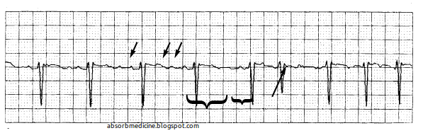
Atrial fibrillation. The tracing demonstrates the absence of P waves (long arrow), as well as the presence of the fine f waves of atrial fibrillation (short arrows). Note the irregularity of the ventricular response, as seen from the variable R-R interval (brackets). [Source]
Common cardiac and non-cardiac causes of aftial fibrillation.

It is not always possible to restore and maintain sinus rhythm in patients with AF.

If sinus rhythm cannot be maintained, treatment should be directed towards controlling the heart rate with
  • digoxin, 
  • ß-blockers, 
  • rate-limiting calcium-channel blockers (verapamil or diltiazem) or 
  • amiodarone. 

Signs of complete heart block

The atrial rate is approximately 100 bpm. The ventricular rate is approximately 40 bpm. The two rates are independent; there is no evidence that any of the atrial impulses are conducted to the ventricles. [Source]
Two ECG strips obtained in the same patient simultaneously (each represents a different vector of the heart’s electrical conduction), demonstrating complete AV block (also called 3rd degree heart block). The solid arrows point to P waves, representing atrial electrical conduction originating from the sinus node. The dashed arrows denote electrical conduction in the ventricles (QRS complexes). Note that the P waves are not related to the QRS complexes, demonstrating that the atria are electrically disconnected from the ventricles. The QRS complexes represent an escape rhythm arising from the ventricle. [Source]

Epsilon Waves in Arrhythmogenic Right Ventricular Dysplasia

The epsilon potential is a right ventricular conduction delay, and appears as a sharp deflection after termination of the QRS complex during the ST segment or upstroke of the T wave. It is seen in the right ventricular leads V1 and V2. (Fontaine named the waves ‘epsilon’ since epsilon follows delta in the
Greek alphabet.)

Causes and ECG Changes in Hypokalaemia

Hypokalaemia is caused by potassium-wasting diuretics, potassium-wasting diarrhoea and hypokalaemic periodic paralysis.

The hallmark of the effect of hypokalaemia on the ECG is the development of large U waves (positive deflection after the T wave). The normal U wave is produced by repolarisation of the His–Purkinje system.

ECG changes in uraemia

A prolonged QT interval is due to hypocalcaemia and tall T waves to hyperkalaemia and/or acidosis, which can be caused by uraemia. The main ECG change resulting from hypocalcaemia is a long QT interval due to prolongation of the ST segment.